Human Gene CACNA1F (ENST00000376265.2) from GENCODE V44
Description: Homo sapiens calcium voltage-gated channel subunit alpha1 F (CACNA1F), transcript variant 1, mRNA. (from RefSeq NM_005183) RefSeq Summary (NM_005183): This gene encodes a multipass transmembrane protein that functions as an alpha-1 subunit of the voltage-dependent calcium channel, which mediates the influx of calcium ions into the cell. The encoded protein forms a complex of alpha-1, alpha-2/delta, beta, and gamma subunits in a 1:1:1:1 ratio. Mutations in this gene can cause X-linked eye disorders, including congenital stationary night blindness type 2A, cone-rod dystropy, and Aland Island eye disease. Alternatively spliced transcript variants encoding multiple isoforms have been observed. [provided by RefSeq, Aug 2013]. Gencode Transcript: ENST00000376265.2 Gencode Gene: ENSG00000102001.13 Transcript (Including UTRs) Position: hg38 chrX:49,205,063-49,233,371 Size: 28,309 Total Exon Count: 48 Strand: - Coding Region Position: hg38 chrX:49,205,137-49,233,309 Size: 28,173 Coding Exon Count: 48
ID:CAC1F_HUMAN DESCRIPTION: RecName: Full=Voltage-dependent L-type calcium channel subunit alpha-1F; AltName: Full=Voltage-gated calcium channel subunit alpha Cav1.4; FUNCTION: Voltage-sensitive calcium channels (VSCC) mediate the entry of calcium ions into excitable cells and are also involved in a variety of calcium-dependent processes, including muscle contraction, hormone or neurotransmitter release, gene expression, cell motility, cell division and cell death. The isoform alpha-1F gives rise to L-type calcium currents. Long-lasting (L-type) calcium channels belong to the 'high-voltage activated' (HVA) group. They are blocked by dihydropyridines (DHP), phenylalkylamines, benzothiazepines, and by omega-agatoxin-IIIA (omega-Aga-IIIA). They are however insensitive to omega-conotoxin- GVIA (omega-CTx-GVIA) and omega-agatoxin-IVA (omega-Aga-IVA). SUBUNIT: Voltage-dependent calcium channels are multisubunit complexes, consisting of alpha-1, alpha-2, beta and delta subunits in a 1:1:1:1 ratio. The channel activity is directed by the pore- forming and voltage-sensitive alpha-1 subunit. In many cases, this subunit is sufficient to generate voltage-sensitive calcium channel activity. The auxiliary subunits beta and alpha-2/delta linked by a disulfide bridge regulate the channel activity. Interacts (via IQ domain) with CABP4; in a calcium independent manner (By similarity). SUBCELLULAR LOCATION: Membrane; Multi-pass membrane protein. TISSUE SPECIFICITY: Expression in skeletal muscle and retina. DOMAIN: Each of the four internal repeats contains five hydrophobic transmembrane segments (S1, S2, S3, S5, S6) and one positively charged transmembrane segment (S4). S4 segments probably represent the voltage-sensor and are characterized by a series of positively charged amino acids at every third position. DISEASE: Defects in CACNA1F are the cause of congenital stationary night blindness type 2A (CSNB2A) [MIM:300071]. Congenital stationary night blindness is a non-progressive retinal disorder characterized by impaired night vision. DISEASE: Defects in CACNA1F are the cause of cone-rod dystrophy X- linked type 3 (CORDX3) [MIM:300476]. CORDs are inherited retinal dystrophies belonging to the group of pigmentary retinopathies. CORDs are characterized by retinal pigment deposits visible on fundus examination, predominantly in the macular region, and initial loss of cone photoreceptors followed by rod degeneration. This leads to decreased visual acuity and sensitivity in the central visual field, followed by loss of peripheral vision. Severe loss of vision occurs earlier than in retinitis pigmentosa. DISEASE: Defects in CACNA1F are the cause of Aaland island eye disease (AIED) [MIM:300600]; also known as Forsius-Eriksson type ocular albinism. On the Aaland island in the Baltic Sea, AIED is an X-linked recessive retinal disease characterized by a combination of fundus hypopigmentation, decreased visual acuity due to foveal hypoplasia, nystagmus, astigmatism, protan color vision defect, myopia, and defective dark adaptation. Except for progression of axial myopia, the disease can be considered to be a stationary condition. Electroretinography reveals abnormalities in both photopic and scotopic functions. SIMILARITY: Belongs to the calcium channel alpha-1 subunit (TC 1.A.1.11) family. CACNA1F subfamily. SEQUENCE CAUTION: Sequence=AAB92359.1; Type=Erroneous gene model prediction; WEB RESOURCE: Name=Mutations of the CCNA1F gene; Note=Retina International's Scientific Newsletter; URL="http://www.retina-international.org/files/sci-news/cacnamut.htm";
The RNAfold program from the Vienna RNA Package is used to perform the secondary structure predictions and folding calculations. The estimated folding energy is in kcal/mol. The more negative the energy, the more secondary structure the RNA is likely to have.
Pfam Domains: PF08763 - Voltage gated calcium channel IQ domain PF00520 - Ion transport protein
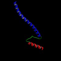
ModBase Predicted Comparative 3D Structure on O60840
Front
Top
Side
The pictures above may be empty if there is no ModBase structure for the protein. The ModBase structure frequently covers just a fragment of the protein. You may be asked to log onto ModBase the first time you click on the pictures. It is simplest after logging in to just click on the picture again to get to the specific info on that model.
Orthologous Genes in Other Species
Orthologies between human, mouse, and rat are computed by taking the best BLASTP hit, and filtering out non-syntenic hits. For more distant species reciprocal-best BLASTP hits are used. Note that the absence of an ortholog in the table below may reflect incomplete annotations in the other species rather than a true absence of the orthologous gene.
Gene Ontology (GO) Annotations with Structured Vocabulary
Molecular Function: GO:0005216 ion channel activity GO:0005244 voltage-gated ion channel activity GO:0005245 voltage-gated calcium channel activity GO:0005262 calcium channel activity GO:0005515 protein binding GO:0008331 high voltage-gated calcium channel activity GO:0046872 metal ion binding
Biological Process: GO:0006811 ion transport GO:0006816 calcium ion transport GO:0007601 visual perception GO:0034765 regulation of ion transmembrane transport GO:0043029 T cell homeostasis GO:0050856 regulation of T cell receptor signaling pathway GO:0050896 response to stimulus GO:0050908 detection of light stimulus involved in visual perception GO:0055085 transmembrane transport GO:0070588 calcium ion transmembrane transport GO:0086010 membrane depolarization during action potential GO:1901386 negative regulation of voltage-gated calcium channel activity